Microestrutura e ultraestrutura do vitelário do inseto cochonilha, Dactylopius coccus (Hemiptera: Dactylopiidae)
Autores
-
Arturo Ramírez-Cruz
Departamento de Investigación. CIIDIR Unidad Michoacán. Instituto Politécnico Nacional. México
 https://orcid.org/0000-0001-5184-6194
DOI:
 https://doi.org/10.30550/j.azl/2404
Palavras-chave:
bactérias simbióticas, corpos fibrogranulares, corpos lamelares, lamínulas anulares, vitelogêneseResumo
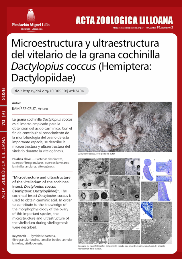
O inseto cochonilha, Dactylopius coccus, é utilizado para a obtenção de ácido carmínico. Para contribuir com a compreensão da morfofisiologia do ovário dessa importante espécie, descrevemos a microestrutura e a ultraestrutura do vitelário durante a vitelogênese. Seus ovaríolos telotróficos são constituídos pelo germário, vitelário e pedicelo. Cada vitelário contém um único folículo ovariano, composto por células foliculares e oócito, e o folículo é revestido externamente por uma túnica própria. As células foliculares eram colunares a cuboides, mononucleadas e continham abundantes ribossomos e mitocôndrias, embora com pouco retículo endoplasmático rugoso. O oócito continha um grande número de mitocôndrias, ribossomos e microtúbulos, mas pouco retículo endoplasmático rugoso e complexos de Golgi. O citoplasma cortical do oócito exibia lamelas anulares, formadas a partir de um corpo fibrogranular. A gema era composta principalmente de grânulos proteicos e glóbulos lipídicos, sendo as proteínas as primeiras a serem incorporadas endocitadamente no oócito. Dentro de suas membranas endocíticas, o material proteico se compacta até a completa formação dos grânulos de gema. Corpos lamelares foram observados próximos a esses grânulos ou suas membranas, sugerindo seu papel na reciclagem dessas membranas. Os oócitos continham bactérias simbióticas, que são transferidas do germário para o oócito através do cordão trófico, pelo menos desde o início da vitelogênese. O estudo de algumas das características microestruturais e ultraestruturais do vitelário, e até mesmo do germário, é importante para o estabelecimento de relações filogenéticas entre os diferentes grupos de Coccomorpha.
Downloads
Referências
Aquino Pérez, G., Bárcenas Ortega, N. M. & Valdez Carrasco, J. (2002). Avances en la citología de la cochinilla del nopal en condiciones in vivo e in vitro y su biología reproductiva. En Portillo, L. & Vigueras, A. L. (Eds.). Memoria del II Congreso Internacional de Grana Cochinilla y Colorantes Naturales y II Reunión Internacional del Grupo de Trabajo en Cochinilla, Cactusnet-FAO (pp. 3-25). Guadalajara: Universidad de Guadalajara.
Berry, S. J. (1985). RNA synthesis and storage during insect oogenesis. In Browder, L. W. (Ed.). Developmental biology. A comprehensive synthesis. Oogenesis (Vol. 1, pp. 351-384). New York: Plenum Press. https://doi.org/10.1007/978-1-4615-6814-8
Bilinski, S. M., Jaglarz, M. K. & Tworzydlo, W. (2017). The pole (germ) plasm in insect oocytes. In Kloc, M. (Ed.). Oocytes. Maternal information and functions. Results and problems in cell differentiation (Vol. 63, pp. 103-126). Switzerland: Springer International Publishing AG. https://doi.org/10.1007/978-3-319-60855-6_5
Bogolyubov, D. & Parfenov, V. (2008). Structure of the insect oocyte nucleus with special reference to interchromatin granule clusters and Cajal bodies. International Review of Cell and Molecular Biology, 269, 59-110. https//doi.org./10.1016/S1937-6448(08)01002-2
Bozzola, J. J. & Russell, L. D. (1999). Electron microscopy. Principles and techniques for biologist (2nd ed.). Boston: Jones and Bartlett Publishers.
Brac, T. (1983). Lamellar bodies are the intracelullar site of membrane turnover in insect fat body. Tissue and Cell, 15(6), 873-884. https://doi.org/10.1016/0040-8166(83)90055-1
Büning, J. (1994). The Insect Ovary: Ultrastructure, previtellogenic growth and evolution. London: Chapman and Hall. https://doi.org/10.1007/978-94-011-0741-9
Chapman, R. F. (2013). The Insects: Structure and function (5th ed.). Simpson, S. J. & Douglas, A. E. (Eds.). New York: Cambridge University Press.
https://doi.org/10.1017/CBO9781139035460
Chu, H. M., Norris, D. M. & Rao, K. D. P. (1982). Sorbic acid-induced differences in the ultrastructural development of oocytes in the microbially ectosymbiotic female of Xyleborus ferrugineus (Fabr.) (Coleoptera, Scolytidae). Journal of Morphology, 173, 313- 324. https://doi.org/10.1002/jmor.1051730308
Conn, D. B. & Quinn, C. M. (1995). Ultrastructure of the vitellogenic egg chambers of the caddisfly Brachycentrus incanus (Insecta: Trichoptera). Invertebrate Biology, 114(4), 334-343. https://doi.org/10.2307/3226842
Flores-Alatorre, H. L., Abrego-Reyes, V., Reyes-Esparza, J. A., Ángeles, E. & Alva-Hurtado, F. (2014). Variation in the concentration of carminic acid produced by Dactylopius coccus (Hemiptera: Dactylopiidae) at various maturation stages. Journal of Economic Entomology, 107(4), 1700-1705.
https://doi.org/10.1603/EC13475
Foldi, I. (1990). Gametogenesis. In Rosen, D. (Ed.). Armored scale insects. Their biology, natural enemies and control (Vol. 4A, pp. 199-204). Netherlands: Elsevier Science.
Foldi, I. (1997). Internal anatomy of the adult female. In Ben-Dov, Y. & Hodgson, C. J. (Eds.). Soft scale insects. Their biology, natural enemies and control (Vol. 7A, pp.73-90). Netherlands: Elsevier Science. https://doi.org/10.1016/S1572-4379(97)80045-0
Gavrilov-Zimin, I. A. (2018). Ontogenesis, morphology and higher classification of archaeococcids (Homoptera: Coccinea: Orthezioidea). Zoosystematica Rossica, Suplementum 2, 1-260. https://doi.org/10.31610/zsr/2018.supl.2.1
Halkka, L. & Halkka, O. (1977). Accumulation of gene products in the oocytes of the dragonfly Cordulia aenea. II. Induction of annulate lamellae within dense masses during diapause. Journal of Cell Science, 26(1), 217-228. https://doi.org/10.1242/jcs.26.1.217
Hopkings, C. R. & King, P. E. (1966). An electron-microscopical and histochemical study of the oocyte periphery in Bombus terrestris during vitellogenesis. Journal of Cell Science, 1, 201-216. https://doi.org/10.1242/jcs.1.2.201
Huebner, E. & Anderson, E. (1972). A cytological study of the ovary of Rhodnius prolixus. II. Oocyte differentiation. Journal of Morphology, 137(4), 385-415.
https://doi.org/10.1002/jmor.1051370402
Huebner, E. (1984). The ultrastructure and development of the telotrophic ovary. In King, R. C. & Akai, H. (Eds.). Insect Ultrastructure (Vol. 2, pp. 3-48). New York: Plenum Press. https://doi.org/10.1007/978-1-4613-2715-8_1
Irles, P., Ramos, S. & Piulachs, M.-D. (2017). SPARC preserves follicular epithelium integrity in insect ovaries. Developmental Biology, 422(2), 105-114.
https://doi.org/10.1016/j.ydbio.2017.01.005
Jedrzejowska, I. & Kubrakiewicz, J. (2004). Ovariole development in telotrophic ovaries of snake flies (Raphidioptera). Folia Biologica (Kraków), 52(3-4), 175-184. https://doi.org/10.3409/1734916044527467
Kessel, R. G. (1981). Origin, differentiation, distribution and possible functional role of annulate lamellae during spermatogenesis in Drosophila melanogaster. Journal of Ultrastructure Research, 75, 72-96.
https://doi.org/10.1016/S0022-5320(81)80101-3
Kessel, R. G. (1983). Fibrogranular bodies, annulate lamellae, and polyribosomes in the dragonfly oocyte. Journal of Morphology, 176, 171-180.
https://doi.org/10.1002/jmor.1051760206
Kessel, R. G. (1985). The relationships of annulate lamellae, fibrogranular bodies, nucleolus, and polyribosomes during spermatogenesis in Drosophila melanogaster. Journal of Ultrastructure Research, 91, 183-191.
https://doi.org/10.1016/S0022-5320(85)80012-5
Kessel, R. G. (1989). The annulate lamellae-from obscurity to spotlight. Electron Microscopy Reviews, 2, 257-348. https://doi.org/10.1016/0892-0354(89)90003-8
Kessel, R. G. (1992). Annulate lamellae: A last frontier in cellular organelles. International Review of Cytology, 133, 43-120. https://doi.org/10.1016/S0074-7696(08)61858-6
King, R.C. & Büning, J. (1985). The origin and functioning of insect oocytes and nurse cells. In Kerkut, G. A. & Gilbert, L. I. (Eds). Comprensive insect physiology, biochemistry and pharmacology (Vol. 1, pp. 37-82). Oxford: Pergamon Press.
Koteja, J., Pyka-Fo?ciak, G., Vogelgesang, M. & Szklarzewicz, T. (2003). Structure of the ovary in Steingelia (Sternorrhyncha: Coccinea), and its phylogenetic implications. Arthropod Structure and Development, 32, 247-256. https://doi.org/10.1016/S1467-8039(03)00046-X
Kunkel, J. G. (1991). Models of pattern formation in insect oocytes. In Vivo, 5, 443-456.
Kurata, S.i., Koga, K. & Sakaguchi, B. (1978). Nucleolar size in parallel with ribosomal RNA synthesis at diapause termination in the eggs of Bombyx mori. Chromosoma (Berl.), 68, 313-317. https://doi.org/10.1007/BF00327166
Lau, Y. T., Reynhout, J. K. & Horowitz, S. B. (1984). Regional water changes during oocyte meiotic maturation: Evidence of ooplasmic segregation. Developmental Biology, 104, 106-110. https://doi.org/10.1016/0012-1606(84)90040-X
Liu, W., Xie, Y., Dong, J., Xue, J., Tian, F. & Wu, J. (2014). Ultra- and microstructure of the female reproductive system of Matsucoccus matsumurae. Arthropod Structure and Development, 43(3), 243-253.
http://doi.org/10.1016/j.asd.2014.02.001
Locke, M. (2003). Surface membranes, Golgi complexes, and vacuolar systems. Annual Review of Entomology, 48, 1-27.
https://doi.org/10.1146/annurev.ento.48.091801.112543
Ma, P. W. K., Baird, S. & Ramaswamy, S. B. (2002). Morphology and formation of the eggshell in the tarnished plant bug, Lygus lineolaris (Palisot de Beauvois) (Hemiptera: Miridae). Arthropod Structure and Development, 31, 131-146.
Mahowald, A. P. (1972). Oogenesis. In Counce, S. J. & Waddington, C. H. (Eds.). Developmental System: Insects (Vol. 1, pp. 1-47). London: Academic Press Inc.
Margaritis, L. H. (1985). Structure and physiology of eggsell. In Kerkut, G. A. & Gilbert L. I. (Eds.). Comprehensive insect physiology, biochemistry and pharmacology (Vol. 1, pp: 153-230). Oxford: Pergamon Press.
Méndez-Gallegos, S. de J., Panzavolta, T. & Tiberi, R. (2003). Carmine cochineal Dactylopius coccus Costa (Rhynchota: Dactylopiidae): Significance, production and use. Advances in Horticultural Science, 17(3), 165-171.
Merisko, E. M. (1989). Annulate lamellae: an organelle in search of a function. Tissue and Cell, 21(3), 343-354. https://doi.org/10.1016/0040-8166(89)90049-9
Michalik, A., Golas, A., Kot, M., Wieczorek, K. & Szklarzewicz, T. (2013). Endosymbiotic microorganisms in Adelges (Sacchiphantes) viridis (Insecta, Hemiptera, Adelgoidea: Adelgidae): Molecular characterization, ultrastructure and transovarial transmission. Arthropod Structure and Development, 42, 531-538. https://doi.org/10.1016/j.asd.2013.09.004
Michalik, K., Szklarzewicz, T., Kalandyk-Kolodziejczyk, M. & Michalik, A. (2019). Bacterial associates of Orthezia urticae, Matsucoccus pini, and Steingelia gorodetskia- scale insects of archaeoccoid families Ortheziidae, Matsucoccidae, and Steingeliidae (Hemiptera, Coccomorpha). Protoplasma, 256, 1205-1215.
https://doi.org/10.1007/s00709-019-01377-z
Ogorzalek, A. (2007). Structural and functional diversification of follicular epithelium in Coreus marginatus (Coreidae: Heteroptera). Arthropod Structure and Development, 36, 209-2019. https://doi.org/10.1016/j.asd.2006.09.004
Raccaud-Schoeller, J. (1980). Les Insectes. Physiologie, développement. Paris: Masson.
Raikhel, A. S. & Dhadialla, T. S. (1992). Accumulation of yolk proteins in insect oocytes. Annual Review of Entomology, 37, 217-251.
https://doi.org/10.1146/annurev.en.37.010192.001245
Ramírez-Cruz, A., Llanderal-Cázares, C. & Racotta, R. (2008). Ovariole structure of the cochineal scale insect, Dactylopius coccus. Journal of Insect Science, 8(20), 1-5. https://doi.org/10.1673/031.008.2001
Ramírez-Cruz, A. (2012). Ultraestructura del germario de Dactylopius coccus Costa, 1829 (Hemiptera: Coccoidea: Dactylopiidae). Acta Zoológica Mexicana (n.s), 28(3), 586-593. https://doi.org/10.21829/azm.2012.283860
Ramírez-Cruz, A. (2014). Maduración de la ovariola de la grana cochinilla Dactylopius coccus (Hemiptera: Coccoidea: Dactylopidae). Revista Colombiana de Entomología, 40(2), 225-229.
Ramírez-Cruz, A. & Llanderal-Cázares, C. (2019). Effect of mating on ovary maturation in Dactylopius coccus (Hemiptera: Coccoidea: Dactylopiidae). Revista Colombiana de Entomología, 45(2), e7954.
https://doi.org/10.25100/socolen.v45i2.7954
Ren, H., Xin, G., Jia, M., Zhu, S., Lin, Q., Wang, X., Jiang, Q. & Zhang, C. (2019). Postmitotic annulate lamellae assembly contributes to nuclear envelope reconstitution in daughter cells. Journal of Biological Chemistry, 294 (27), 10383-10391. https://doi.org/10.1074/jbc.ac119.008171
Rosenblueth, M., Martínez-Romero, J., Ramírez-Puebla, S. T., Vera-Ponce de León, A., Rosas-Pérez, T., Bustamante-Brito, R., Rincón-Rosales, R. & Martínez-Romero, E. (2018). Endosymbiotic microorganisms of scale insects. TIP Revista Especializada en Ciencias Químico-Biológicas, 21(1), 53-69.
https://doi.org/10.1016/j.recqb.2017.08.006
Schmitz, G. & Müller, G. (1991). Structure and function of lamellar bodies, lipid-protein complexes involved in storage and secretion of cellular lipids. Journal of Lipid Research, 32, 1539-1570. https://doi.org/10.1016/S0022-2275(20)41642-6
Snigirevskaya, E. S., Hays, A. R. & Raikhel, A. S. (1997). Secretory and internalization pathways of mosquito yolk protein precursors. Cell and Tissue Research, 290, 129-142. https://doi.org/10.1007/s004410050915
Stebbings, H., Lane, J. D. & Talbot, N. J. (1995). mRNA translocation and microtubules: insect ovary models. Trends in Cell Biology, 5, 361-365.
https://doi.org/10.1016/S0962-8924(00)89070-4
Szklarzewicz, T., Kedra, K. & Niznik, S. (2005). Ultrastructural studies of the ovary of Palaeococcus fuscipennis (Burmeister) (Insecta, Hemiptera, Coccinea: Monophlebidae). Folia Biologica (Kraków), 53 (1-2), 45-50. https://doi.org/10.3409/1734916054663483
Szklarzewicz, T., Kedra, K., & Niznik, S. (2006). Ultrastructure and transovarial transmission of endosymbiotic microorganism in Palaeococcus fuscipennis (Burmeister) (Insecta, Hemiptera, Coccinea: Monophlebidae). Folia Biologica (Kraków), 54(1-2), 69-74.
http://doi.org/10.3409/173491606777919102
Szklarzewicz, T., Jankowska, W., Lukasiewicz, K. & Szymanska, B. (2007). Structure of the ovaries and oogenesis in Cixius nervosus (Cixiidae), Javesella pellucida and Conomelus anceps (Delphacidae) (Insecta, Hemiptera, Fulgoromorpha). Arthropod Structure and Development, 36, 199-207.
https://doi.org/10.1016/j.asd.2006.09.001
Szklarzewicz, T., Kalandyk-Kolodziejczyk, M., Kot, M. & Michalik, A. (2013). Ovary structure and transovarial transmission of endosymbiotic microorganisms in Marchalina hellenica (Insecta, Hemiptera, Coccomorpha: Marchalinidae). Acta Zoologica (Stockholm), 94, 184-192.
https://doi.org/10.1111/j.1463-6395.2011.00538.x
Szklarzewicz, T., Michalik, A., Kalandyk-Kolodziejczyk, M., Kobialka, M. & Simon, E. (2014). Ovary of Matsucoccus pini (Insecta, Hemiptera, Coccinea: Matsucoccidae): Morphology, ultrastructure, and phylogenetic implications. Microscopy Research and Technique, 77, 327-334.
https://doi.org/10.1002/jemt.22347
Szklarzewicz, T., Michalik, A. & Michalik, K. (2020). The diversity of symbiotic systems in scale insects. In Kloc, M. (Ed.). Simbiosis: Cellular, molecular, medical and evolutionary aspects. Results and problems in cell differentiation (Vol. 69, pp. 469-495). Switzerland: Springer Nature Switzerland AG. https://doi.org/10.1007/978-3-030-51849-3_18
Tamarelle, M. (1979). Recherches ultrastructurales sur la segregation et le developpement de la lignee germinale chez les embryons de quatre collemboles (Insecta: Apterygota). International Journal of Insect Morphology and Embryology, 8(2), 95-111. https://doi.org/10.1016/0020-7322(79)90009-6
Tremblay, E. (1997). Endosymbiosis. In Ben-Dov, Y. & Hodgson, C. J. (Eds.). Soft scale insects. Their biology, natural enemies and control (Vol. 7A, pp. 261-267). Netherlands: Elsevier Science. https://doi.org/10.1016/S1572-4379(97)80056-5
Williams, D. J. & Hodgson, C. J. (2014). The case for using the infraorder Coccomorpha above the superfamily Coccoidea for the scale insects (Hemiptera: Sternorrhyncha). Zootaxa, 3869 (3), 348-350.
Downloads
Publicado
Como Citar
Edição
Seção
Licença
Copyright (c) 2026 Acta Zoológica Lilloana

Este trabalho está licenciado sob uma licença Creative Commons Attribution-NonCommercial-NoDerivatives 4.0 International License.