Microestructura y ultraestructura del vitelario de la grana cochinilla Dactylopius coccus (Hemiptera: Dactylopiidae)
Autores/as
-
Arturo Ramírez-Cruz
Departamento de Investigación. CIIDIR Unidad Michoacán. Instituto Politécnico Nacional. México
 https://orcid.org/0000-0001-5184-6194
DOI:
 https://doi.org/10.30550/j.azl/2404
Palabras clave:
bacterias simbiontes, cuerpos fibrogranulares, cuerpos lamelares, laminillas anulares, vitelogénesisResumen
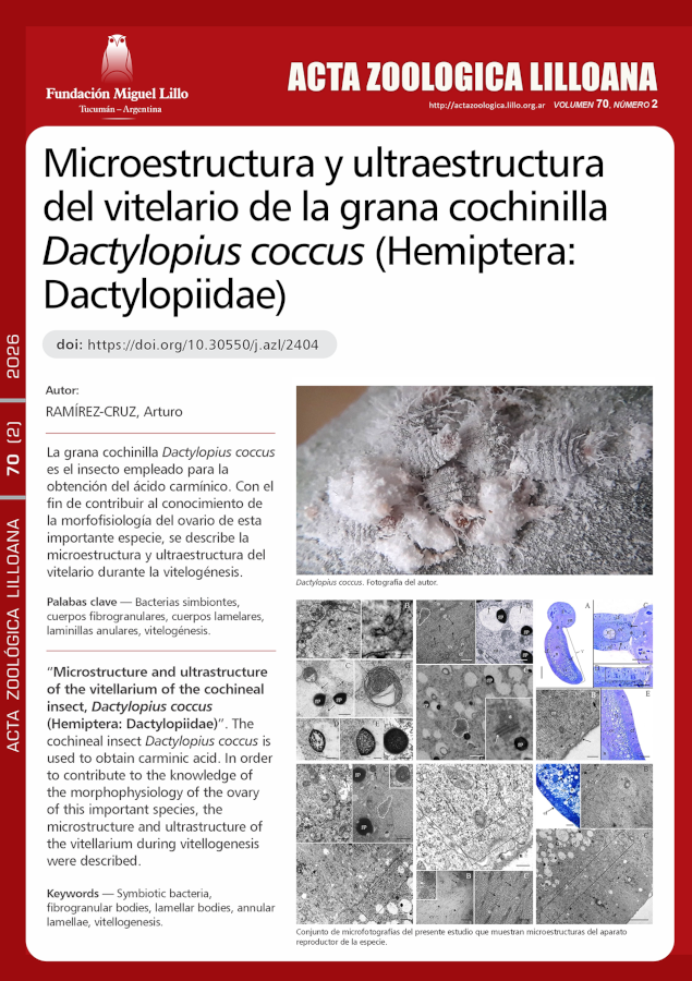
La grana cochinilla Dactylopius coccus es el insecto empleado para la obtención del ácido carmínico. Con el fin de contribuir al conocimiento de la morfofisiología del ovario de esta importante especie, se describe la microestructura y ultraestructura del vitelario durante la vitelogénesis. Sus ovariolas de tipo telotrófico, están formadas por el germario, el vitelario y el pedicelo. Cada vitelario contiene un solo folículo ovárico, constituido por las células foliculares y el ovocito. El folículo externamente está cubierto de una túnica propia. Las células foliculares fueron entre columnares y cúbicas, mononucleadas y con abundantes ribosomas y mitocondrias, aunque escaso retículo endoplásmico rugoso. El ovocito presentó gran cantidad de mitocondrias, ribosomas, microtúbulos, pero
escasos retículo endoplásmico rugoso y complejo de Golgi. El citoplasma cortical del ovocito presentó laminillas anulares, formadas a partir de un cuerpo fibrogranular. El vitelo se formó principalmente de gránulos de proteínas y de glóbulos de lípidos, siendo las proteínas las primeras
en incorporarse endocíticamente al ovocito; dentro de sus membranas endocíticas el material proteínico se compacta hasta la formación completa de gránulos de vitelo. Próximos a estos gránulos o sus membranas se observaron cuerpos lamelares, sugiriendo que éstos participan reciclando dichas membranas. Los ovocitos presentaron bacterias simbiontes, que son transferidas desde el germario hacia el ovocito, mediante el cordón trófico, al menos desde el inicio de la vitelogénesis.
Descargas
Citas
Aquino Pérez, G., Bárcenas Ortega, N. M. & Valdez Carrasco, J. (2002). Avances en la citología de la cochinilla del nopal en condiciones in vivo e in vitro y su biología reproductiva. En Portillo, L. & Vigueras, A. L. (Eds.). Memoria del II Congreso Internacional de Grana Cochinilla y Colorantes Naturales y II Reunión Internacional del Grupo de Trabajo en Cochinilla, Cactusnet-FAO (pp. 3-25). Guadalajara: Universidad de Guadalajara.
Berry, S. J. (1985). RNA synthesis and storage during insect oogenesis. In Browder, L. W. (Ed.). Developmental biology. A comprehensive synthesis. Oogenesis (Vol. 1, pp. 351-384). New York: Plenum Press. https://doi.org/10.1007/978-1-4615-6814-8
Bilinski, S. M., Jaglarz, M. K. & Tworzydlo, W. (2017). The pole (germ) plasm in insect oocytes. In Kloc, M. (Ed.). Oocytes. Maternal information and functions. Results and problems in cell differentiation (Vol. 63, pp. 103-126). Switzerland: Springer International Publishing AG. https://doi.org/10.1007/978-3-319-60855-6_5
Bogolyubov, D. & Parfenov, V. (2008). Structure of the insect oocyte nucleus with special reference to interchromatin granule clusters and Cajal bodies. International Review of Cell and Molecular Biology, 269, 59-110. https//doi.org./10.1016/S1937-6448(08)01002-2
Bozzola, J. J. & Russell, L. D. (1999). Electron microscopy. Principles and techniques for biologist (2nd ed.). Boston: Jones and Bartlett Publishers.
Brac, T. (1983). Lamellar bodies are the intracelullar site of membrane turnover in insect fat body. Tissue and Cell, 15(6), 873-884. https://doi.org/10.1016/0040-8166(83)90055-1
Büning, J. (1994). The Insect Ovary: Ultrastructure, previtellogenic growth and evolution. London: Chapman and Hall. https://doi.org/10.1007/978-94-011-0741-9
Chapman, R. F. (2013). The Insects: Structure and function (5th ed.). Simpson, S. J. & Douglas, A. E. (Eds.). New York: Cambridge University Press.
https://doi.org/10.1017/CBO9781139035460
Chu, H. M., Norris, D. M. & Rao, K. D. P. (1982). Sorbic acid-induced differences in the ultrastructural development of oocytes in the microbially ectosymbiotic female of Xyleborus ferrugineus (Fabr.) (Coleoptera, Scolytidae). Journal of Morphology, 173, 313- 324. https://doi.org/10.1002/jmor.1051730308
Conn, D. B. & Quinn, C. M. (1995). Ultrastructure of the vitellogenic egg chambers of the caddisfly Brachycentrus incanus (Insecta: Trichoptera). Invertebrate Biology, 114(4), 334-343. https://doi.org/10.2307/3226842
Flores-Alatorre, H. L., Abrego-Reyes, V., Reyes-Esparza, J. A., Ángeles, E. & Alva-Hurtado, F. (2014). Variation in the concentration of carminic acid produced by Dactylopius coccus (Hemiptera: Dactylopiidae) at various maturation stages. Journal of Economic Entomology, 107(4), 1700-1705.
https://doi.org/10.1603/EC13475
Foldi, I. (1990). Gametogenesis. In Rosen, D. (Ed.). Armored scale insects. Their biology, natural enemies and control (Vol. 4A, pp. 199-204). Netherlands: Elsevier Science.
Foldi, I. (1997). Internal anatomy of the adult female. In Ben-Dov, Y. & Hodgson, C. J. (Eds.). Soft scale insects. Their biology, natural enemies and control (Vol. 7A, pp.73-90). Netherlands: Elsevier Science. https://doi.org/10.1016/S1572-4379(97)80045-0
Gavrilov-Zimin, I. A. (2018). Ontogenesis, morphology and higher classification of archaeococcids (Homoptera: Coccinea: Orthezioidea). Zoosystematica Rossica, Suplementum 2, 1-260. https://doi.org/10.31610/zsr/2018.supl.2.1
Halkka, L. & Halkka, O. (1977). Accumulation of gene products in the oocytes of the dragonfly Cordulia aenea. II. Induction of annulate lamellae within dense masses during diapause. Journal of Cell Science, 26(1), 217-228. https://doi.org/10.1242/jcs.26.1.217
Hopkings, C. R. & King, P. E. (1966). An electron-microscopical and histochemical study of the oocyte periphery in Bombus terrestris during vitellogenesis. Journal of Cell Science, 1, 201-216. https://doi.org/10.1242/jcs.1.2.201
Huebner, E. & Anderson, E. (1972). A cytological study of the ovary of Rhodnius prolixus. II. Oocyte differentiation. Journal of Morphology, 137(4), 385-415.
https://doi.org/10.1002/jmor.1051370402
Huebner, E. (1984). The ultrastructure and development of the telotrophic ovary. In King, R. C. & Akai, H. (Eds.). Insect Ultrastructure (Vol. 2, pp. 3-48). New York: Plenum Press. https://doi.org/10.1007/978-1-4613-2715-8_1
Irles, P., Ramos, S. & Piulachs, M.-D. (2017). SPARC preserves follicular epithelium integrity in insect ovaries. Developmental Biology, 422(2), 105-114.
https://doi.org/10.1016/j.ydbio.2017.01.005
Jedrzejowska, I. & Kubrakiewicz, J. (2004). Ovariole development in telotrophic ovaries of snake flies (Raphidioptera). Folia Biologica (Kraków), 52(3-4), 175-184. https://doi.org/10.3409/1734916044527467
Kessel, R. G. (1981). Origin, differentiation, distribution and possible functional role of annulate lamellae during spermatogenesis in Drosophila melanogaster. Journal of Ultrastructure Research, 75, 72-96.
https://doi.org/10.1016/S0022-5320(81)80101-3
Kessel, R. G. (1983). Fibrogranular bodies, annulate lamellae, and polyribosomes in the dragonfly oocyte. Journal of Morphology, 176, 171-180.
https://doi.org/10.1002/jmor.1051760206
Kessel, R. G. (1985). The relationships of annulate lamellae, fibrogranular bodies, nucleolus, and polyribosomes during spermatogenesis in Drosophila melanogaster. Journal of Ultrastructure Research, 91, 183-191.
https://doi.org/10.1016/S0022-5320(85)80012-5
Kessel, R. G. (1989). The annulate lamellae-from obscurity to spotlight. Electron Microscopy Reviews, 2, 257-348. https://doi.org/10.1016/0892-0354(89)90003-8
Kessel, R. G. (1992). Annulate lamellae: A last frontier in cellular organelles. International Review of Cytology, 133, 43-120. https://doi.org/10.1016/S0074-7696(08)61858-6
King, R.C. & Büning, J. (1985). The origin and functioning of insect oocytes and nurse cells. In Kerkut, G. A. & Gilbert, L. I. (Eds). Comprensive insect physiology, biochemistry and pharmacology (Vol. 1, pp. 37-82). Oxford: Pergamon Press.
Koteja, J., Pyka-Fo?ciak, G., Vogelgesang, M. & Szklarzewicz, T. (2003). Structure of the ovary in Steingelia (Sternorrhyncha: Coccinea), and its phylogenetic implications. Arthropod Structure and Development, 32, 247-256. https://doi.org/10.1016/S1467-8039(03)00046-X
Kunkel, J. G. (1991). Models of pattern formation in insect oocytes. In Vivo, 5, 443-456.
Kurata, S.i., Koga, K. & Sakaguchi, B. (1978). Nucleolar size in parallel with ribosomal RNA synthesis at diapause termination in the eggs of Bombyx mori. Chromosoma (Berl.), 68, 313-317. https://doi.org/10.1007/BF00327166
Lau, Y. T., Reynhout, J. K. & Horowitz, S. B. (1984). Regional water changes during oocyte meiotic maturation: Evidence of ooplasmic segregation. Developmental Biology, 104, 106-110. https://doi.org/10.1016/0012-1606(84)90040-X
Liu, W., Xie, Y., Dong, J., Xue, J., Tian, F. & Wu, J. (2014). Ultra- and microstructure of the female reproductive system of Matsucoccus matsumurae. Arthropod Structure and Development, 43(3), 243-253.
http://doi.org/10.1016/j.asd.2014.02.001
Locke, M. (2003). Surface membranes, Golgi complexes, and vacuolar systems. Annual Review of Entomology, 48, 1-27.
https://doi.org/10.1146/annurev.ento.48.091801.112543
Ma, P. W. K., Baird, S. & Ramaswamy, S. B. (2002). Morphology and formation of the eggshell in the tarnished plant bug, Lygus lineolaris (Palisot de Beauvois) (Hemiptera: Miridae). Arthropod Structure and Development, 31, 131-146.
Mahowald, A. P. (1972). Oogenesis. In Counce, S. J. & Waddington, C. H. (Eds.). Developmental System: Insects (Vol. 1, pp. 1-47). London: Academic Press Inc.
Margaritis, L. H. (1985). Structure and physiology of eggsell. In Kerkut, G. A. & Gilbert L. I. (Eds.). Comprehensive insect physiology, biochemistry and pharmacology (Vol. 1, pp: 153-230). Oxford: Pergamon Press.
Méndez-Gallegos, S. de J., Panzavolta, T. & Tiberi, R. (2003). Carmine cochineal Dactylopius coccus Costa (Rhynchota: Dactylopiidae): Significance, production and use. Advances in Horticultural Science, 17(3), 165-171.
Merisko, E. M. (1989). Annulate lamellae: an organelle in search of a function. Tissue and Cell, 21(3), 343-354. https://doi.org/10.1016/0040-8166(89)90049-9
Michalik, A., Golas, A., Kot, M., Wieczorek, K. & Szklarzewicz, T. (2013). Endosymbiotic microorganisms in Adelges (Sacchiphantes) viridis (Insecta, Hemiptera, Adelgoidea: Adelgidae): Molecular characterization, ultrastructure and transovarial transmission. Arthropod Structure and Development, 42, 531-538. https://doi.org/10.1016/j.asd.2013.09.004
Michalik, K., Szklarzewicz, T., Kalandyk-Kolodziejczyk, M. & Michalik, A. (2019). Bacterial associates of Orthezia urticae, Matsucoccus pini, and Steingelia gorodetskia- scale insects of archaeoccoid families Ortheziidae, Matsucoccidae, and Steingeliidae (Hemiptera, Coccomorpha). Protoplasma, 256, 1205-1215.
https://doi.org/10.1007/s00709-019-01377-z
Ogorzalek, A. (2007). Structural and functional diversification of follicular epithelium in Coreus marginatus (Coreidae: Heteroptera). Arthropod Structure and Development, 36, 209-2019. https://doi.org/10.1016/j.asd.2006.09.004
Raccaud-Schoeller, J. (1980). Les Insectes. Physiologie, développement. Paris: Masson.
Raikhel, A. S. & Dhadialla, T. S. (1992). Accumulation of yolk proteins in insect oocytes. Annual Review of Entomology, 37, 217-251.
https://doi.org/10.1146/annurev.en.37.010192.001245
Ramírez-Cruz, A., Llanderal-Cázares, C. & Racotta, R. (2008). Ovariole structure of the cochineal scale insect, Dactylopius coccus. Journal of Insect Science, 8(20), 1-5. https://doi.org/10.1673/031.008.2001
Ramírez-Cruz, A. (2012). Ultraestructura del germario de Dactylopius coccus Costa, 1829 (Hemiptera: Coccoidea: Dactylopiidae). Acta Zoológica Mexicana (n.s), 28(3), 586-593. https://doi.org/10.21829/azm.2012.283860
Ramírez-Cruz, A. (2014). Maduración de la ovariola de la grana cochinilla Dactylopius coccus (Hemiptera: Coccoidea: Dactylopidae). Revista Colombiana de Entomología, 40(2), 225-229.
Ramírez-Cruz, A. & Llanderal-Cázares, C. (2019). Effect of mating on ovary maturation in Dactylopius coccus (Hemiptera: Coccoidea: Dactylopiidae). Revista Colombiana de Entomología, 45(2), e7954.
https://doi.org/10.25100/socolen.v45i2.7954
Ren, H., Xin, G., Jia, M., Zhu, S., Lin, Q., Wang, X., Jiang, Q. & Zhang, C. (2019). Postmitotic annulate lamellae assembly contributes to nuclear envelope reconstitution in daughter cells. Journal of Biological Chemistry, 294 (27), 10383-10391. https://doi.org/10.1074/jbc.ac119.008171
Rosenblueth, M., Martínez-Romero, J., Ramírez-Puebla, S. T., Vera-Ponce de León, A., Rosas-Pérez, T., Bustamante-Brito, R., Rincón-Rosales, R. & Martínez-Romero, E. (2018). Endosymbiotic microorganisms of scale insects. TIP Revista Especializada en Ciencias Químico-Biológicas, 21(1), 53-69.
https://doi.org/10.1016/j.recqb.2017.08.006
Schmitz, G. & Müller, G. (1991). Structure and function of lamellar bodies, lipid-protein complexes involved in storage and secretion of cellular lipids. Journal of Lipid Research, 32, 1539-1570. https://doi.org/10.1016/S0022-2275(20)41642-6
Snigirevskaya, E. S., Hays, A. R. & Raikhel, A. S. (1997). Secretory and internalization pathways of mosquito yolk protein precursors. Cell and Tissue Research, 290, 129-142. https://doi.org/10.1007/s004410050915
Stebbings, H., Lane, J. D. & Talbot, N. J. (1995). mRNA translocation and microtubules: insect ovary models. Trends in Cell Biology, 5, 361-365.
https://doi.org/10.1016/S0962-8924(00)89070-4
Szklarzewicz, T., Kedra, K. & Niznik, S. (2005). Ultrastructural studies of the ovary of Palaeococcus fuscipennis (Burmeister) (Insecta, Hemiptera, Coccinea: Monophlebidae). Folia Biologica (Kraków), 53 (1-2), 45-50. https://doi.org/10.3409/1734916054663483
Szklarzewicz, T., Kedra, K., & Niznik, S. (2006). Ultrastructure and transovarial transmission of endosymbiotic microorganism in Palaeococcus fuscipennis (Burmeister) (Insecta, Hemiptera, Coccinea: Monophlebidae). Folia Biologica (Kraków), 54(1-2), 69-74.
http://doi.org/10.3409/173491606777919102
Szklarzewicz, T., Jankowska, W., Lukasiewicz, K. & Szymanska, B. (2007). Structure of the ovaries and oogenesis in Cixius nervosus (Cixiidae), Javesella pellucida and Conomelus anceps (Delphacidae) (Insecta, Hemiptera, Fulgoromorpha). Arthropod Structure and Development, 36, 199-207.
https://doi.org/10.1016/j.asd.2006.09.001
Szklarzewicz, T., Kalandyk-Kolodziejczyk, M., Kot, M. & Michalik, A. (2013). Ovary structure and transovarial transmission of endosymbiotic microorganisms in Marchalina hellenica (Insecta, Hemiptera, Coccomorpha: Marchalinidae). Acta Zoologica (Stockholm), 94, 184-192.
https://doi.org/10.1111/j.1463-6395.2011.00538.x
Szklarzewicz, T., Michalik, A., Kalandyk-Kolodziejczyk, M., Kobialka, M. & Simon, E. (2014). Ovary of Matsucoccus pini (Insecta, Hemiptera, Coccinea: Matsucoccidae): Morphology, ultrastructure, and phylogenetic implications. Microscopy Research and Technique, 77, 327-334.
https://doi.org/10.1002/jemt.22347
Szklarzewicz, T., Michalik, A. & Michalik, K. (2020). The diversity of symbiotic systems in scale insects. In Kloc, M. (Ed.). Simbiosis: Cellular, molecular, medical and evolutionary aspects. Results and problems in cell differentiation (Vol. 69, pp. 469-495). Switzerland: Springer Nature Switzerland AG. https://doi.org/10.1007/978-3-030-51849-3_18
Tamarelle, M. (1979). Recherches ultrastructurales sur la segregation et le developpement de la lignee germinale chez les embryons de quatre collemboles (Insecta: Apterygota). International Journal of Insect Morphology and Embryology, 8(2), 95-111. https://doi.org/10.1016/0020-7322(79)90009-6
Tremblay, E. (1997). Endosymbiosis. In Ben-Dov, Y. & Hodgson, C. J. (Eds.). Soft scale insects. Their biology, natural enemies and control (Vol. 7A, pp. 261-267). Netherlands: Elsevier Science. https://doi.org/10.1016/S1572-4379(97)80056-5
Williams, D. J. & Hodgson, C. J. (2014). The case for using the infraorder Coccomorpha above the superfamily Coccoidea for the scale insects (Hemiptera: Sternorrhyncha). Zootaxa, 3869 (3), 348-350.
Descargas
Publicado
Cómo citar
Número
Sección
Licencia
Derechos de autor 2026 Acta Zoológica Lilloana

Esta obra está bajo una licencia internacional Creative Commons Atribución-NoComercial-SinDerivadas 4.0.