Histomorfología e histoquímica del tubo digestivo en hembras de Leptodactylus latinasus (Anura: Leptodactylidae) durante el periodo reproductivo
Autores/as
-
Eliana Elizabeth Heredia Ojeda
Centro de Referencia para Lactobacilos (CERELA-CONICET)
 https://orcid.org/0009-0009-2103-0675
-
Franco José Pucci Alcaide
Instituto de Morfología Animal, Dirección Zoología
https://orcid.org/0000-0001-6509-7331
-
Adriana Azucena Michel
Instituto de Morfología Animal, Dirección Zoología, Fundación Miguel Lillo
https://orcid.org/0000-0002-5799-7003
-
Ana Pucci Alcaide
Cátedra de Histología, Facultad de Ciencias Naturales e Inst. Miguel Lillo, Universidad Nacional de Tucumán
https://orcid.org/0009-0000-2725-4391
DOI:
Palabras clave:
Histología, Digestión, Glándulas gástricas, ReproducciónResumen
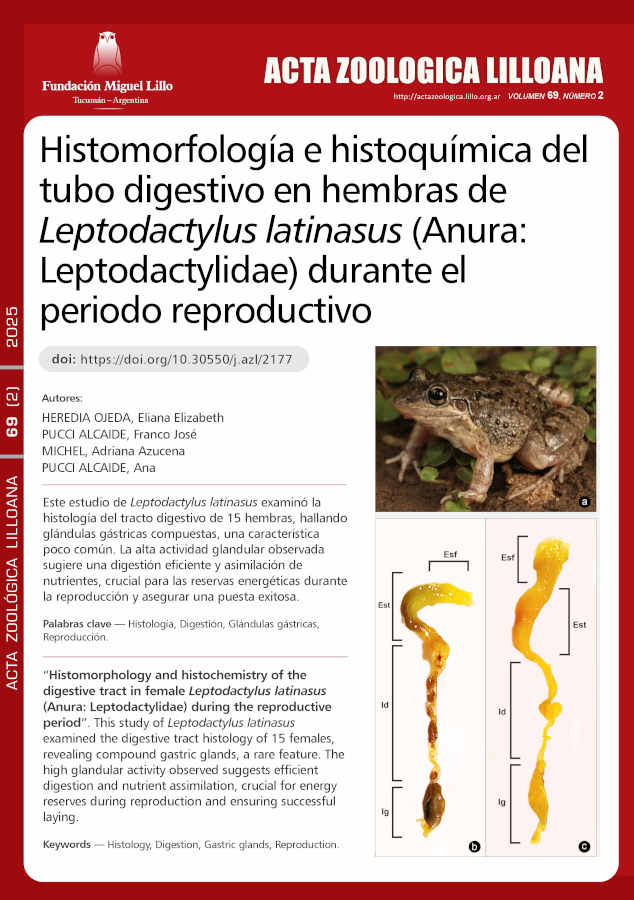
El tubo digestivo metaboliza los alimentos necesarios para el aporte de energía. El estado funcional en el que se encuentran los órganos que lo conforman representa una asociación clara entre el hábitat y la fisiología del animal. El objetivo es analizar histológicamente las estructuras célulo-tisulares del esófago, estómago, intestino delgado y grueso, a fin de aportar datos a la comprensión integral del sistema y su efecto directo sobre la biología reproductiva de la especie. Se diseccionaron 15 ejemplares hembra de Leptodactylus latinasus, los órganos fueron removidos, limpiados y procesados con la técnica histológica de rutina, los cortes se colorearon con Hematoxilina Eosina, Tricrómico de Mallory; Alcian Blue a pH 0,5 y 2,5- Ácido Periódico Schiff. El análisis histomorfológico reveló la presencia de características compartidas con otras especies, sin embargo, la presencia de glándulas gástricas compuestas por: 1) células mucosas del cuello, 2) células principales y 3) células parietales, parece ser un atributo no compartido por la mayoría de las especies. Por otra parte, en el análisis histoquímico se describen seis tipos de glándulas que se diferencian de acuerdo a la naturaleza química de su contenido. Éste estudio demuestra que los ejemplares hembra de Leptodactylus latinasus presentan características histológicas a lo largo del tubo digestivo que le permiten una eficiente digestión y asimilación del alimento, la elevada actividad glandular observada mediante el análisis histoquímico refleja un momento de actividad en el proceso de alimentación durante el periodo reproductivo asegurando reservas energéticas que permitirán una puesta exitosa.
Descargas
Citas
Akat, E., Ar?kan H. & Göçmen B. (2014). Histochemical and Biometric Study of the Gastrointestinal System of Hyla orientalis (Anura, Hylidae) (Bedriaga, 1890). European Journal of Histochemistry. 58 (4): 2452. DOI: https://doi.org/10.4081/ejh.2014.2452
Antoniazzi, C. E., Llanes, R. E., & Armando, A. P. (2017). Características de los anfibios. Universidad Nacional del Litoral. 45-53.
Bernarde, P. S. & Anjos, L. D. (1999). Distribuição espacial e temporal da anurofauna no Parque Estadual Mata dos Godoy, Londrina, Paraná, Brasil (Amphibia: Anura). Comunicações do Museu de Ciências e Tecnologia PUCRS. Série Zoologia. Vol. 12: 127-140.
Birdsey, G. M., Lewin, J., Holbrook, J. D., Simpson, V. R., Cunningham, A. A. & Danpure, C. J. (2005). A comparative analysis of the evolutionary relationship between diet and enzyme targeting in bats, marsupials and other mammals. Proceedings of the Royal Society B: Biological Sciences. 272 (1565): 833-840. DOI: https://doi.org/10.1098/rspb.2004.3011
Castro, J. C, Lima, S. L., Braga, G. T., Azevedo, R. V., Pinto, C. E. L. & Silva, A. R. (2010). Anatomo-histologia do esôfago da rã touro (Lithobates catesbeianus SHAW, 1802). PUBVET, Publicações em Medicina Veterinária e Zootecnia. Vol. 4, N. 41, Ed. 146, Art. 980.
Derting, T.L. & Bogue, B.A. (1993). Responses of the gut to moderate energy demands in a small herbivore (Microtus pennsylvanicus). Journal of Mammalogy, Volume 74, Issue 1, Pages 59–68. DOI: https://doi.org/10.2307/1381905
Duellman, W.E., & Trueb, L. (1986). Biology of Amphibians. The Johns Hopkins University Press, Maryland, 670 pp. DOI: https://doi.org/10.2307/1445022
Ferri, D., Liquori, G. E., Natale, L., Santarelli, G., & Scillitani, G. (2001). Mucin histochemistry of the digestive tract of the red-legged frog Rana aurora aurora. Acta Histochemica. Volume 103, Issue 2, Pages 225-237. DOI: https://doi.org/10.1078/0065-1281-00582
Flores, E. E. & Aranzábal, M. D. C. U. (2002). Atlas de histología de vertebrados. UNAM. Pág. 71-90.
Gallardo, J. M. (1964). “Leptodactylus prognathus” Boul. & “L. mystacinus” (Burm.) con sus respectivas especies aliadas. Revista del Museo Argentino de Ciencias Naturales Bernardino Rivadavia, Instituto Nacional de Investigaciones en Ciencias Naturales (Argentina) Zoología, 9: 91-121.
Gallego-Huidobro J & Pastor LM. (1996). Histology of the mucosa of the oesophagogastric junction and the stomach in adult Rana perezi. Journal of Anatomy. Vol. 188 (Pt 2): 439-444.
Geuze, J.J. (1971). Light and electron microscope observation on the gastric mucosa of the frog (Rana esculenta): Zeitschrift für Zellforschung und Mikroskopische Anatomie. Vol. 117, pages 87-102. DOI: https://doi.org/10.1007/BF00331104
Giaretta A. A. & Kokubum M. (2004). Reproductive ecology of Leptodactylus furnarius Sazima and Bokermann, 1978, a frog that lays eggs in underground chambers. Herpetozoa 16(3/4): 115–126.
Green D. A. & Millar J. S. (1987). Changes in gut dimensions and capacity of Peromyscus maniculatus relative to diet quality and energy needs. Canadian Journal of Zoology 65: 2159-2162. DOI: https://doi.org/10.1139/z87-329
Gross J. E., Wang Z. & Wunder B. A. (1985). Effects of food quality and energy needs: changes in gut morphology and capacity of Microtus ochrogaster. Journal of Mammalogy Vol. 66. Issue 4: 661-667. DOI: https://doi.org/10.2307/1380792
Heyer, W. R. (1969a). The adaptive ecology of the species groups of the genus Leptodactylus (Amphibia, Leptodactylidae). Evolution. Vol. 23, No. 3, pp. 421-428. DOI: https://doi.org/10.1111/j.1558-5646.1969.tb03525.x
Heyer, W. R. (1969b). Studies on the genus Leptodactylus (Amphibia, Leptodactylidae). III. A redefinition of the genus Leptodactylus and a description of a new genus of leptodactylid frogs. Contributions in Science, Los Angeles County Museum of Natural History. Vol. 155: 1–14. DOI: https://doi.org/10.5962/p.241143
Heyer, W. R. (1978). Systematics of the Fuscus group of the frog genus Leptodactylus (Amphibia, Leptodactylidae). Natural History Museum of Los Angeles County, Science Bulletin. Vol. 29: 1–85. DOI: https://doi.org/10.5479/si.00810282.301
Hirji, K. N. & Nikundiwe, A. M. (1982). Observations on the oesophageal glands in some Tanzanian anurans. Department of Zoology and Marine Biology, University of Dar es Salaam. Vol. 17: 32-34. DOI: https://doi.org/10.1080/02541858.1982.11447775
Jiménez de la Espada, M. (1875). Vertebrados del Viaje al Pacífico Verificado de 1862 a 1865 por una Comisión de Naturalistas Enviada por el Gobierno Español. Batracios. Madrid: A. Miguel Ginesta. DOI: https://doi.org/10.5962/bhl.title.5769
Lavilla E. O., Heyer, R. W., Kwet, A., & Langone, J. (2004). Leptodactylus latinasus. The IUCN Red List of Threatened Species 2004: e. T57139A11590252.
Lavilla, E. O. (2018). Reproducción y desarrollo en anuros argentinos. Sistemática y Filogenia de los Vertebrados, 3ra edición. 222-227.
Liquori, G. E., Scillitani, G., Mastrodonato, M., & Ferri, D. (2002). Histochemical investigations on the secretory cells in the oesophagogastric tract of the Eurasian green toad, Bufo viridis. The Histochemical Journal. Vol. 34: 517-524. DOI: https://doi.org/10.1023/A:1024766124211
Lopez-Calleja M. V., Bozinovic F. & Martínez del Río C. (1997). Effects of sugar concentration on hummingbird feeding and energy use. Comparative and Biochemical Physiology. Vol. 118: 1291-1299. DOI: https://doi.org/10.1016/S0300-9629(97)00243-0
Naya D, Bozinovic F & P Sabat. (2008). Ecología nutricional y flexibilidad digestiva en anfibios. In: Herpetología en Chile, (M. Vidal & A. Labra, eds.), Capítulo XV, Science-Verlag. pp. 427-447.
Machado-Santos C., Pelli-Martins A. A., Abidu-Figueiredo M. & de Brito-Gitirana L. (2014). Histochemical and immunohistochemical analysis of the stomach of Rhinella icterica (Anura, Bufonidae). Journal of Histology. Article ID 872795, 8 pages. DOI: https://doi.org/10.1155/2014/872795
Martori, R.; Aun L. & Rocha C. (1999). Variación estacional de la dieta de Liolaemus wiegmanni (Squamata: Tropiduridae) en un agroecosistema del sur de Córdoba, Argentina. Cuadernos de Herpetología. Vol. 13. Nº 1-2: 69-80.
Martori, R.; Juárez, R. & Aun, L. (2002). La taxocenosis de lagartos de Achiras, Córdoba, Argentina: parámetros biológicos y estado de conservación. Revista Española de Herpetología. Vol. 16: 73-91.
McWilliams, S.R. & Karasov, W.H. (2001). Phenotypic flexibility in digestive system structure and function in migratory birds and its ecological significance. Comparative Biochemistry and Physiology. Vol. 128(3): 579-593. DOI: https://doi.org/10.1016/S1095-6433(00)00336-6
Medina R. G., Ponssa, M. L. & Aráoz, E. (2016). Environmental, land cover and land use constraints on the distributional patterns of anurans: Leptodactylus species (Anura, Leptodactylidae) from Dry Chaco. PeerJ Inc. 1-27. DOI: https://doi.org/10.7717/peerj.2605
Menin M. & Giaretta A. A. (2003). Predation on foam nests of Leptodactyline frogs (Anura: Leptodactylidae) by larvae of Beckeriella niger (Diptera: Ephydridae). Journal of Zoology. Vol. 261: 239–243. DOI: https://doi.org/10.1017/S0952836903004138
Moss R. (1974). Winter diets, gut lengths, and interespecific competition in Alaskan Ptarmigan. Vol. 91: 737-746. DOI: https://doi.org/10.2307/4084726
Ponssa M. L & J. S. Barrionuevo. (2008). Foam-generating behavior in tadpoles of Leptodactylus latinasus (Amphibia, Leptodactylidae): significance in systematics. Zootaxa 1884: 51–59. DOI: https://doi.org/10.11646/zootaxa.1884.1.3
Ponssa, M. L., Medina, R. G. & Vera Candiotti, M. F. (2019). Ranita silbadora Leptodactylus latinasus. Universo Tucumano. Fundación Miguel Lillo. N°33. 1-15.
Pucci, A., Ponssa, M. L., Pucci, F., & Alcaide, M. (2012). Histología de ovario en hembras vitelogénicas de Leptodactylus latinasus (Anura, Leptodactylidae). Acta zoológica lilloana. 56 (1-2): 44-53.
Pucci, A., Pucci, F. J., & Alcaide, M. F. (2016). Histología e histoquímica del oviducto de Leptodactylus latinasus (Anura: Leptodactylidae). Período reproductivo preovulatorio. Acta zoológica lilloana. 60 (1): 68-77.
Pucci, A., Pucci, F. J., Michel, A. A., & Ponssa, M. L. (2020). Testicular histology of Anurans that deposit eggs out of water. Acta zoológica lilloana. 64 (2): 84-115. DOI: https://doi.org/10.30550/j.azl/2020.64.2/2
Santana, M. A. & Menin, E. (1997). Histologia de esôfago de Leptodactylus labyrinthicus Spix, 1824 (Amphibia, Anura, Leptodactylidae). Ceres, 44(251): 111-123.
Secor, S.M. & Diamond, J.M. (2000). Evolution of regulatory responses to feeding in snakes. Physiological and Biochemical Zoology. Vol. 73(2): 123-141. DOI: https://doi.org/10.1086/316734
Secor, S. M. (2001). Regulation of digestive performance: a proposed adaptive response. Comparative Biochemistry and Physiology Part A: Vol 128. 565-577. DOI: https://doi.org/10.1016/S1095-6433(00)00325-1
Secor, S. M. (2005). Evolutionary and cellular mechanisms regulating intestinal performance of amphibians and reptiles. Integrative and Comparative Biology. Vol. 45: 282-294. DOI: https://doi.org/10.1093/icb/45.2.282
Sibly R. M. (1981). Estrategias de digestión y defecación. En: Townsend C. R., Calow P (eds) Ecología fisiológica: un enfoque evolutivo del uso de los recursos. Blackwell, Oxford. 109-139.
Starck, J. M. (1999). Structural flexibility of the gastro-intestinal tract of vertebrates - implications for evolutionary morphology. Zoologischer Anzeiger. Vol 238(1-2): 87-101.
Starck, J. M. (2003). Shaping up: how vertebrates adjust their digestive system to changing environmental conditions. Animal Biology. Vol. 53(3): 245-257. DOI: https://doi.org/10.1163/157075603322539444
Stevens, C. E., & Hume, I. D. (1995). Comparative physiology of the vertebrate digestive system, second ed. Cambridge University Press, Cambridge.
Vaira M., Akmentins, M., Attademo, M., Baldo, D., Barrasso, D., Barrionuevo, J. S., Basso, N., Blotto, B., Cairo, S., Cajade, R., Céspedez J., Corbalán, V., Chilote, P. Duré M., Falcione C., Ferraro D., Gutiérrez F. R., Ingaramo M. R., Junges C., Lajmanovich R., Lescano J. N., Marangoni F., Martinazzo L., Marti R., Moreno L., Natale G. S., Pérez Iglesia J. M., Peltze P., Quiroga L., Rosset S., Sanabria E., Sánchez L., Schaefer E., Úbeda C. & Zaracho, V. (2012). Categorización del estado de conservación de los anuros de la República Argentina. Cuadernos de Herpetología. Vol. 26 supl 1.1: 131–159.
Valverde, B. S., Fanali, L. Z., Franco-Belussi, L. & de Oliveira, C. (2019). Comparative morphology of the digestive tract of two Neotropical tree frogs (Genus Boana). Zoologischer Anzeiger. Vol. 281: 44-52. DOI: https://doi.org/10.1016/j.jcz.2019.05.002
Vitt, L. J. & Caldwell, J. P. (2014). Herpetology: an introductory biology of amphibians and reptiles. Fourth ed. Academic Press. DOI: https://doi.org/10.1016/B978-0-12-386919-7.00002-2
Descargas
Publicado
Cómo citar
Número
Sección
Licencia
Derechos de autor 2025 Acta Zoológica Lilloana

Esta obra está bajo una licencia internacional Creative Commons Atribución-NoComercial-SinDerivadas 4.0.