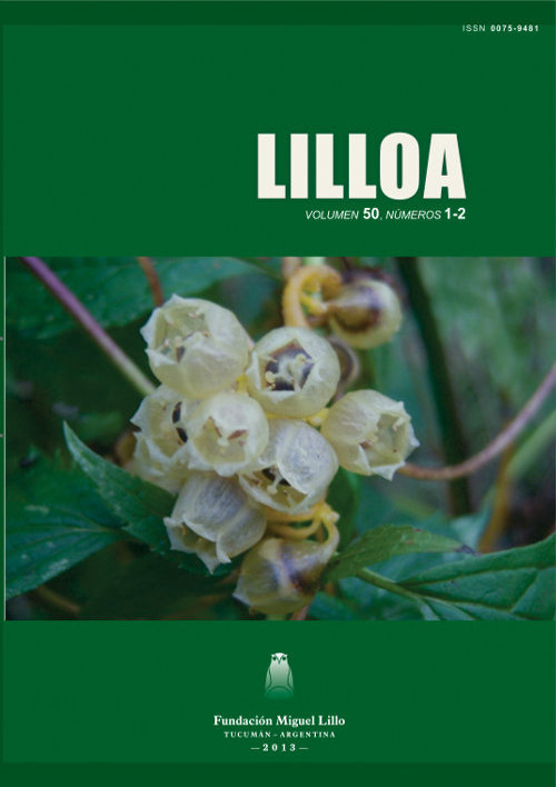
Leaf morpho-anatomy and foliar architecture of Carica quercifolia (Caricaceae)
Keywords:
Carica, colleters, laticifers, leafAbstract
Ruiz, Ana I.; María E. Guantay; María I. Mercado; Graciela Ponessa. 2013. Lilloa 50 (2). “Leaf morpho-anatomy and foliar architecture of Carica quercifolia (Caricaceae)”. The present work aims to describe the morpho-anatomy, foliar architecture and diagnostic characters of a population of Carica quercifolia (A. St.-Hil.) Hieron., located in Villa Nougues, Tucuman (Argentina). Plant material was fixed in FAA and glutaraldehyde. Conventional histological techniques were used for ulterior observations by optical and scanning electron microscopy. C. quercifolia presents foliar dimorphism. Simple, pinnati-lobed, ovate to oblong leaves. Dark green adaxial surface presents glandular areas, of colleters associated with eglandular trichomes, located at the leaf base on primary and secondary veins. The abaxial surface presents light green color with prominent veins. The leaf is hipostomatic, with dorsiventral mesophyll and papillose abaxial epidermal cells. Pinnate, camptodromous, brochidodromous venation. The middle vein is formed by parenchyma pith surrounded by collateral vascular bundles with different maturity degree. Anomocytic, actinocytic and hemiparacytic stomata. Articulated anastomosing laticifers and short stalk colleters with a conoidal axis were observed. Petiole sub-circular to sub-triangular with collateral vascular bundles. Presence of colleters and the foliar architecture of C. quercifolia are described for the first time. The diagnostic characters observed were: foliar architecture, colleters, stomata and trichome types.
Downloads
References
Arambarri, A. M.; S. E. Freire; M. Colares; N. D. Bayón; M. C. Novoa; C. Monti y S. M. Hernández. 2009. Micrografía foliar de arbustos y pequeños árboles medicinales de la Provincia Biogeográfica de las Yungas (Argentina). Kurtziana 35 (1): 15-45.
Barboza, G.E.; J.J. Cantero; C. Núñez; A. Pacciaroni y L.A., Espinar. 2009. Medicinal plants: A general review and a phytochemical and ethnopharmacological screening of the native Argentine Flora. Kurtziana 34 (1-2): 7-365.
Bianchi, A.R. y C. Yáñez. 2005. Las precipitaciones en el Noroeste argentino. En: A. Bianchi; C. Yáñez y L. Acuña. Base de datos mensuales de precipitaciones del Noroeste argentino. Instituto Nacional de Tecnología Agropecuaria (INTA)(editor). Centro Regional Salta-Jujuy, 41pp.
D’Ambrogio de Argüeso, A. 1986. Manual de Técnicas en Histología Vegetal. Editora Hemisferio Sur S. A., Buenos Aires, 83 pp.
Dilcher, D.L. 1974. Approaches to the identification of angiosperm leaves. The Botanical Review 40 (1): 1-157.
Digilio, A.P.L. y P.R. Legname. 1966. Los árboles indígenas de la provincia de Tucumán. Opera Lilloana 15: 20, 129 pp.
Dizeo de Strittmater, C. G. 1973. Nueva técnica de diafanización. Boletín Sociedad Argentina Botánica, 15 (1): 126-129.
Esau, K. 2008. Anatomía vegetal. Editora Omega, Barcelona, España, 641 pp.
Hickey, L. J. 1974. Clasificación de la arquitectura de las hojas de Dicotiledóneas. Boletín Sociedad Argentina Botánica, 16 (1-2): 1-26.
Hickey, L. J. 1979. A revised classification of the architecture of dicotyledonous leaves. En C. R. Metcalfe y L. Chalk (eds.) Anatomy of the Dicotyledons. Volumen I. Second Edition. Editora Clarendon Press, Oxford, 25-39.
Johansen, D.A. 1940. Plant microtechnique. Editora McGraw-Hill. New York, 523 pp.
Metcalfe, C. R. y L. C. Chalk. 1950. Anatomy of the Dicotyledons. Editora Clarendon Press, Oxford: 1145-1156.
Roth, I. 1990. Peculiar surface structures of tropical leaves for gas exchange, guttation, and light capture (Fissures in the epidermis, lenticels, giant stomata, glands, ocelli, papillas, and surface sculpturing). En: B. Rollet, Ch. Högermann y I. Roth (editores), Stratification of Tropical Forest as seen in Leaf Structure (Part 2), 185-238.
Wilkinson, H. 1979. The Plant Surface (Mainly leaf). Part. V. The Cuticle. En: C.R. Metcalfe y L. Chalk (editores), Anatomy of the Dicotyledons. Clarendon, Oxford, Inglaterra, Vol. I: 140-156.
Zuloaga F. O., O. Morrone y M. J. Belgrano. 2008. Catálogo de Plantas Vasculares del Cono Sur (Argentina, Sur de Brasil, Chile, Paraguay y Uruguay). Editora Missouri Botanical Garden, Saint Louis, Missouri, 3348 pp.


